Faculty/Retired
University of Minnesota
Department of Genetics, Cell Biology and Development
6-160 Jackson Hall
321 Church St SE
Minneapolis, MN 55455
Robert L. Sorenson, Ph.D.
Professor Emeritus
University of Minnesota
Department of Genetics, Cell Biology and Development
6-160 Jackson Hall
321 Church St SE
Minneapolis, MN 55455
Download the User Guide v1.1 (PDF) to learn about new platform features.
Each slide is shown with additional information to its right. The image can be changed using any combination of the following commands.
Sidebar
Links: Click to navigate to a specific region
Images: Click to show this view
Toolbar: Use controls to adjust magnification and pan the image
Mouse
Zoom In: Click left button
Zoom Out: Double-click left button
Pan/Move: Click and drag the image
Keyboard
Zoom In: ‘A’ key
Zoom Out: ‘Z’ key
Pan/Move: Arrow keys (Up, Down, Left, Right)
Reset View: ESC key (fit-to-screen view)
Touch
Tap: Zoom in on a specific area
Double-tap: Zoom out from the current view
Drag: Pan the image
SHARE
A link to a virtual slide can be saved for later viewing in different ways.
Clipboard
The address of this view has been copied to your clipboard. This link can be pasted in any other program.
Bookmark
A bookmark link can be created using the bookmark function (Ctrl-D for Windows or Cmd-D for Mac) of your browser. Choose a name for the bookmark and select the folder in which you want it saved.
MH 016 Simple Epithelia
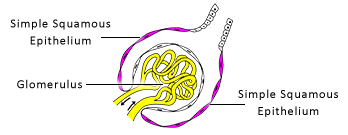
Simple Squamous Epithelium
The contains many different types of epithelia. However, the focus here is only on a simple squamous epithelium.
Find one of the round structures (~250 µm diameter) known as . Each contains a glomerulus (a tuft of capillaries) surrounded by Bowman's capsule.
The interior of the capsule is lined by a that rests on a thick basement membrane. The only part of these cells that can be seen are their nuclei bulging into the interior.